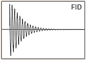
Magnetic Resonance Spectroscopy (MRS) is a non-invasive imaging technique that measure relative levels of tissue metabolites in the form of NMR spectrum. Atoms with an odd mass number such as 1H, 31P and 13C possess the quantum property of “spin” and behave as dipoles aligning along the axis of an applied magnetic field. During relaxation following excitation, radiofrequency signals are generated which can be expressed as a frequency spectrum. Hydrogen is the most abundant atom in living organisms and using high power magnetic fields on in vitro samples, high resolution metabolic spectra can be obtained with clearly defined metabolite peaks of small molecules. Theoretically, MRS can be performed with nuclei of 1H, 23Na, 31P, 13C, But presently for patients 1H (Proton), 31P (Non Proton) spectroscopy is used for clinical evaluation
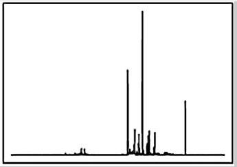
In routine MRI images are formed by signal from water and fat. In MRS the signal from water and fat is suppressed to detect small metabolites. Further the size and shape of peaks depends on the concentration of active nuclei, T1, T2 and T2* effects, splitting by J coupling and overlapping peaks.
Large water peaks are suppressed by using a method in which group of CHESS “Chemical Shift Selective” pulses are tuned to the water resonance to suppress the water.
Inversion Recovery (IR sequences) and Basing eliminate unwanted fat signal to enhance metabolite concentration.
Localization and Pulse Sequence in MR spectroscopy
There are different in vivo MRS sequences have been developed for clinical whole-body MRS, differing in pulse sequences and localization methods. Stimulated echo acquisition mode (STEAM) and point-resolved spectroscopy (PRESS) are commonly used. The signal intensity acquired with PRESS is twice as high as STEAM. Both can be produced with short echo times. The major difference between PRESS and STEAM is the way signal is produced. STEAM uses magnetization to form a stimulated echo, whereas PRESS measures spin echo. Large water peaks must be suppressed in order to visualize low concentration metabolites. Chemical Shift selective (Chess) is used to suppress water. Large amount of fat in the body is problematic to visualize small metabolites; like lipid contamination in breast region or prostate in pelvic fat region, or scalp and marrow fat in Brain. Inversion recovery method and basing eliminates fat signal to visualize metabolic concentration.
Lorem ipsum dolor sit amet, consectetur adipiscing elit. Ut elit tellus, luctus nec ullamcorper mattis, pulvinar dapibus leo.