
MRI Image formation
MRI Knowledge Hub
Magnetic Resonance Imaging (MRI) Basics
MRI is based on the nuclear magnetic resonance (NMR) phenomenon first observed by Bloch and Purcell in 1946. NMR principles have made high quality image recovery possible by allowing the encoding of spatial information into signals that can be detected outside the body. The MRI hardware consists of a main magnet along with three gradient coils to provide smaller magnetic fields. The MRI machine is a large cube with a horizontal tube, known as the bore, running through the center. The patient lies on his back or stomach so that the part of the body to be scanned is at the isocenter of the main magnet. The main magnet provides a very stable and intense magnetic field while the gradient coils provide variable localized fields. Transmitter coils generate oscillating magnetic fields as probe signals while receiver coils convert magnetic fields into electrical signals. NMR is based on the principle that any object can be subdivided into atoms. Each atom is composed of two types of charged particles: protons and electrons. Subatomic particles such as electrons and protons have a property known as spin. Although a proper explanation of subatomic particle behavior requires quantum mechanics, MRI principles can be accurately described using classical models because MRI involves collections of particles. Spin is often associated with the angular momentum of the atom and may be visualized as a top spinning about its vertical axis. Spin values come in integer multiples of 0.5 and may be positive or negative. The spin for an unpaired subatomic particle is 0.5. The net spin of a collection of particles is the sum of the individual spins. Techniques based on NMR, including MRI, are dependent on collections of particles with net spin. According to Ampere’s law, a time-varying electric charge distribution induces a magnetic field. As a consequence, the nucleus of an atom with net spin will produce a small magnetic field. This magnetic field is represented by the vector quantity and known as the nuclear magnetic dipole moment or the magnetic moment. The magnetic moment is related to the spin angular momentum.
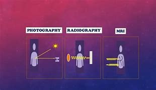
Photography Radiography Magnetic Resonance Imaging
- In photography we illuminate the object and light reflected from the object, falling on sensor or phography paper converted into image.
- Radiography is a technique to take picture inside body by sensing attenuated or reflected beam of EM waves passing inside body.
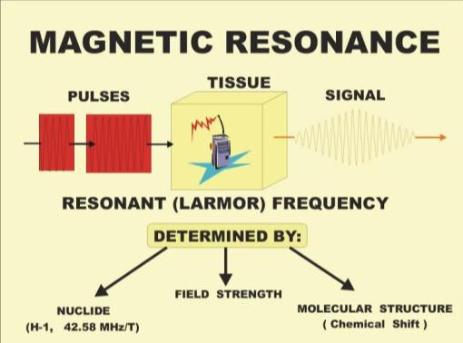
- In Magnetic Resonance Imaging tissue voxels are exited to enegrize by ElectroMagnetic waves in the present of strong Magnetic field then energy released is captured to convert into image.
MRI
To know more on MRI basics and image formation press MRI IMAGE FORMATION