MR Image Processing
A Research and Practical Application based library of Scientific, Technical & Medical Literature
Visualization Segmentation Analysis
MRI Knowledge Hub
Current Approach in MRI Image Processing
Attributes to the World of MR Image processing
The biggest challenge of radiologists, even today, is to have an early diagnosis of diseases from medical images. From the days of Roentgen’s discovery of X-ray in 1895, radiologists are doing the interpretation of medical images. The practice has largely remained unchanged although new modalities of imaging like CT, SPECT, ultrasound and MRI (Magnetic Resonance Imaging) have emerged. There is a lot of research interest in the area for developing new methods and tools for visualization, processing, and analysis of medical images. MR images have received considerable attention because of their additional capability to display changes in the soft tissue.
One of the areas where magnetic resonance imaging is widely used is brain tumor cases and its response to therapy (like radiation therapy and chemotherapy). The image differentiation in tissue within the tumor area (like edema, nacrotic and scar), and early detection of changes in the images due to therapy in the tumor and surrounding regions are important aspects for the diagnosis and management of brain tumor cases. Research workers from different disciplines like computer vision, graphics, image processing, MR physics, medical physics etc. are contributing towards development of MRI based techniques. Efforts are also being made to improve the image quality in order to improve the interpretation of MR image data.
In MR images soft tissue boundaries are visible. However, these boundaries are not identical for different patients. There are variations in shape as well as intensity distribution. Consequently, development of automated tools for analysis of MR images is a challenging and complex problem. Image processing techniques for visualization, segmentation and diagnosis based upon MR Images of brain offers good results. Image processing based applications have been widely used in digital images also. They offer great help in diagnosis and therapy.

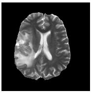
MR Image with soft boundaries

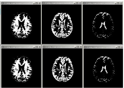
MR Image showing soft segmentation

MR image showing Crisp segmentation
Publications/Patents
Patents
US4430748A: Image thresholding system
US5305204A: Digital image display apparatus with automatic window level and window width adjustment

